Anti-Ksp-Cadherin (Kidney-Specific Cadherin) / CDH16 (CDH16/1532R)
CAT:
37-BNUB1532-500
Size:
500 µL
Price:
Ask
- Availability: 24/48H Stock Items & 2 to 6 Weeks non Stock Items.
- Dry Ice Shipment: No


Anti-Ksp-Cadherin (Kidney-Specific Cadherin) / CDH16 (CDH16/1532R)
Description:
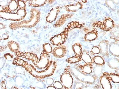
This MAb recognizes a protein of 130 kDa, identified as Ksp-cadherin. Cadherins form a superfamily of related glycoproteins that mediate calcium-dependent cell adhesion and transmit signals from the extracellular matrix to the cytoplasm. Cadherins have been implicated in embryogenesis, tissue morphogenesis, tissue structure maintenance, cell polarization, neoplastic invasiveness and metastasis, and membrane transport. It is suggested that Ksp-cadherin is a marker for terminal differentiation of the basolateral membranes of renal tubular epithelial cells. Within the kidney, Ksp-Cadherin is found exclusively in the basolateral membrane of renal tubular epithelial cells and collecting duct cells, and not in glomeruli, renal interstitial cells, or blood vessels. Ksp-Cadherin has been suggested to distinguish Chromophobe Renal-Cell Carcinoma from Oncocytoma.Primary antibodies are available purified, or with a selection of fluorescent CF® Dyes and other labels. CF® Dyes offer exceptional brightness and photostability. Note: Conjugates of blue fluorescent dyes like CF®405S and CF®405M are not recommended for detecting low abundance targets, because blue dyes have lower fluorescence and can give higher non-specific background than other dye colors.Synonyms:
Cadherin-16 (CDH16); Kidney-specific cadherin; Ksp-cadherin antibodyUNSPSC:
41116161UNSPSC Description:
Primary and secondary antibodies for multiple methodology immunostaining detection applicationGene Name:
CDH16Gene ID:
1014NCBI Gene ID:
513660UniProt:
O75309Cellular Locus:
Plasma membraneHost:
RabbitSpecies Reactivity:
Human, Mouse, RatImmunogen:
Recombinant human full-length CDH16 proteinTarget Antigen:
CDH16 | Ksp-CadherinClonality:
Recombinant MonoclonalIsotype:
IgG κClone:
CDH16/1532RConjugation:
Purified, with BSADisease:
TumorSource:
AnimalApplications:
Flow (verified) | IHC, FFPE (verified) | WB (verified)Validated Applications:
FC, IHC, FFPE, WBField of Research:
Cell adhesionPositive Control:
Normal kidney or renal cell carcinomaConcentration:
0.2 mg/mLBuffer:
PBS, 0.05% BSA, 0.05% azideMolecular Weight:
130 kDaAdditionnal Information:
Higher concentration may be required for direct detection using primary antibody conjugates than for indirect detection with secondary antibody|Immunofluorescence: 1-2 ug/mL|Immunohistology (formalin): 0.5-1 ug/mL|Staining of formalin-fixed tissues requires boiling tissue sections in 10 mM Tris with 1 mM EDTA Buffer pH 9.0 for 10-20 min followed by cooling at RT for 20 min|Flow Cytometry 0.5-1 ug/million cells/0.1 mL|Optimal dilution for a specific application should be determined by userShipping Conditions:
Room temperatureStorage Conditions:
4°C; Stable at room temperature or 37°C (98°F) for 7 days.Shelf Life:
2 yearsCAS Number:
9007-83-4
DATASHEET Document
View DocumentMSDS Document
View Document