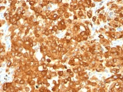
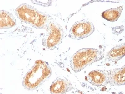
Anti-Pmel17 / gp100 / SILV(HMB45), CF568 conjugate
CAT:
37-BNC680444-100
Size:
100 µL
Price:
Ask
- Availability: 24/48H Stock Items & 2 to 6 Weeks non Stock Items.
- Dry Ice Shipment: No


Anti-Pmel17 / gp100 / SILV(HMB45), CF568 conjugate
Description:
By immunohistochemistry, this antibody specifically recognizes a protein in melanocytes and melanomas. It reacts with junctional and blue nevus cells and variably with fetal and neonatal melanocytes. Intradermal nevi, normal adult melanocytes, and non-melanocytic cells are negative. It does not stain tumor cells of epithelial, lymphoid, glial, or mesenchymal origin. Metastatic amelanotic melanoma can often be confused with a variety of poorly differentiated carcinomas, large cell lymphomas, and sarcomas using H & E stains alone. It is also difficult to differentiate melanoma from spindle cell carcinomas and various types of mesenchymal neoplasms. This MAb stains fetal and neonatal melanocytes, junctional and blue nevus cells, malignant melanoma, and angiomyolipoma (PEComa).Primary antibodies are available purified, or with a selection of fluorescent CF® Dyes and other labels. CF® Dyes offer exceptional brightness and photostability. Note: Conjugates of blue fluorescent dyes like CF®405S and CF®405M are not recommended for detecting low abundance targets, because blue dyes have lower fluorescence and can give higher non-specific background than other dye colors.Synonyms:
95kDa melanocyte-specific secreted glycoprotein, M-beta, Melanocyte lineage specific antigen GP100, Melanocyte protein Pmel 17, Melanoma associated ME20 antigen, Melanosomal matrix protein17, p100, p26, PMEL17, Premelanosome protein, Secreted melanoma-associated ME20 antigen, SILV, Silver homologUNSPSC:
41116161UNSPSC Description:
Primary and secondary antibodies for multiple methodology immunostaining detection applicationGene Name:
PMELGene ID:
6490NCBI Gene ID:
95972UniProt:
P40967Cellular Locus:
Endoplasmic reticulum|Golgi apparatusHost:
MouseSpecies Reactivity:
HumanImmunogen:
Extract of pigmented melanoma metastases from lymph nodesTarget Antigen:
gp100 | Pmel17 | SILVClonality:
MonoclonalIsotype:
IgG1 κClone:
HMB45Conjugation:
CF568Disease:
TumorSource:
AnimalApplications:
IHC, FFPE (verified) | WB (verified)Validated Applications:
IHC, FFPE, WBField of Research:
CancerPositive Control:
SK-MEL-28 cells or MelanomaConcentration:
0.1 mg/mLBuffer:
PBS, 0.1% BSA, 0.05% azideMolecular Weight:
90-100 kDaAdditionnal Information:
Higher concentration may be required for direct detection using primary antibody conjugates than for indirect detection with secondary antibody|Immunofluorescence: 0.5-1 ug/mL|Immunohistology (formalin)|Staining of formalin-fixed tissues requires boiling tissue sections in 10 mM citrate buffer, pH 6.0, for 10-20 min followed by cooling at RT for 20 minutes|Flow Cytometry 0.5-1 ug/million cells/0.1 mL|Does not react with dog or rat, others not tested|Optimal dilution for a specific application should be determined by userShipping Conditions:
Room temperatureStorage Conditions:
4°C; Protect from light; Stable at room temperature or 37°C (98°F) for 7 days.Shelf Life:
2 yearsCAS Number:
9007-83-4
DATASHEET Document
View DocumentMSDS Document
View Document