Anti-Mucin 1 / EMA / Episialin / CD227(GP1.4 + E29), Biotin conjugate
CAT:
37-BNCB0743-100
Size:
100 µL
Price:
Ask
- Availability: 24/48H Stock Items & 2 to 6 Weeks non Stock Items.
- Dry Ice Shipment: No


Anti-Mucin 1 / EMA / Episialin / CD227(GP1.4 + E29), Biotin conjugate
Description:
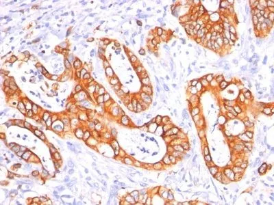
In Western blotting, it recognizes proteins in MW range of 265-400 kDa, identified as different glycoforms of EMA. The α subunit has cell adhesive properties. It can act both as an adhesion and an anti-adhesion protein. EMA may provide a protective layer on epithelial cells against bacterial and enzyme attack. The β subunit contains a C-terminal domain, which is involved in cell signaling, through phosphorylations and protein-protein interactions. In immunohistochemical assays, it superbly stains routine formalin/paraffin carcinoma tissues. Antibody to EMA is useful as a pan-epithelial marker for detecting early metastatic loci of carcinoma in bone marrow or liver.Primary antibodies are available purified, or with a selection of fluorescent CF® Dyes and other labels. CF® Dyes offer exceptional brightness and photostability. Note: Conjugates of blue fluorescent dyes like CF®405S and CF®405M are not recommended for detecting low abundance targets, because blue dyes have lower fluorescence and can give higher non-specific background than other dye colors.Synonyms:
Breast carcinoma-associated antigen DF3; CA15-3; Carcinoma-associated mucin Episialin; Epithelial Membrane Antigen; H23AG; KL-6; MAM6; MUC-1; MUC-1/SEC; MUC-1/X; MUC1-alpha; MUC1-beta; MUC1-CT; MUC1-NT; MUC1/ZD; Mucin 1 cell surface associated; Mucin-1 subunit beta; Peanut-reactive urinary mucin; PEM; PEMT; Polymorphic epithelial mucin; PUM; Tumor-associated epithelial membrane antigenUNSPSC:
41116161UNSPSC Description:
Primary and secondary antibodies for multiple methodology immunostaining detection applicationGene Name:
MUC1Gene ID:
4582NCBI Gene ID:
89603UniProt:
P15941Cellular Locus:
Plasma membraneHost:
MouseSpecies Reactivity:
HumanImmunogen:
Human milk fat globule membranes (GP1.4); Delipidated extract of human milk fat globule membranes (E29)Target Antigen:
CD227 | EMA | Episialin | Mucin 1Clonality:
MonoclonalIsotype:
IgGClone:
GP1.4 E29Conjugation:
BiotinDisease:
TumorSource:
AnimalApplications:
IHC, FFPE (verified)Validated Applications:
IHC, FFPEField of Research:
Cancer, MucinsPositive Control:
MCF-7 or MDA-231 cells. Breast, colon, ovarian, endometrial carcinoma.Concentration:
0.1 mg/mLBuffer:
PBS, 0.1% BSA, 0.05% azideMolecular Weight:
265-400 kDaAdditionnal Information:
Higher concentration may be required for direct detection using primary antibody conjugates than for indirect detection with secondary antibody|Immunofluorescence: 0.5-1 ug/mL|Immunohistology formalin-fixed 0.1-0.2 ug/mL|Staining of formalin-fixed tissues requires boiling tissue sections in 10 mM citrate buffer, pH 6.0, for 10-20 min followed by cooling at RT for 20 minutes|Flow Cytometry 0.5-1 ug/million cells/0.1 mL|Optimal dilution for a specific application should be determined by userShipping Conditions:
Room temperatureStorage Conditions:
4°C; Stable at room temperature or 37°C (98°F) for 7 days.Shelf Life:
2 yearsCAS Number:
9007-83-4
DATASHEET Document
View DocumentMSDS Document
View Document