Anti-Ep-CAM / CD326(VU-1D9)
CAT:
37-BNUM0017-50
Size:
50 µL
Price:
Ask
- Availability: 24/48H Stock Items & 2 to 6 Weeks non Stock Items.
- Dry Ice Shipment: No


Anti-Ep-CAM / CD326(VU-1D9)
Description:
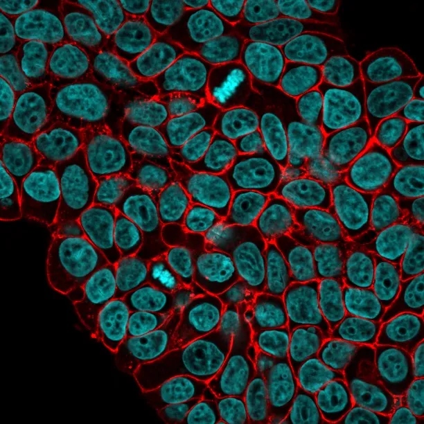
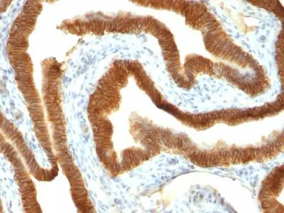
This antibody reacts with the first EGF repeat in the extracellular domain of Ep-CAM. It is a 40-43 kDa transmembrane epithelial glycoprotein, also identified as epithelial specific antigen (ESA), or epithelial cellular adhesion molecule (Ep-CAM). It is expressed on baso-lateral cell surface in most simple epithelia and a vast majority of carcinomas with the exception of adult squamous epithelium, hepatocytes and gastric epithelial cells. This antibody has been used to distinguish adenocarcinoma from pleural mesothelioma and hepatocellular carcinoma. This antibody is also useful in distinguishing serous carcinomas of the ovary from mesothelioma._x000D_ _x000D_ Primary antibodies are available purified, or with a selection of fluorescent CF® Dyes and other labels. CF® Dyes offer exceptional brightness and photostability. Note: Conjugates of blue fluorescent dyes like CF®405S and CF®405M are not recommended for detecting low abundance targets, because blue dyes have lower fluorescence and can give higher non-specific background than other dye colors._x000D_ _x000D_Synonyms:
Adenocarcinoma-associated Antigen; Cell Surface Glycoprotein Trop-1; EGP2; EGP314; EGP40; Epithelial Cell Adhesion Molecule; Epithelial Glycoprotein 314; ESA; KSA; TACD1; TROP1; Tumor-associated Calcium Signal Transducer 1 (TACSTD1)UNSPSC:
41116161UNSPSC Description:
Primary and secondary antibodies for multiple methodology immunostaining detection applicationGene Name:
TACSTD1Gene ID:
4072NCBI Gene ID:
542050UniProt:
P16422Cellular Locus:
Exosomes/EVs|Plasma membraneHost:
MouseSpecies Reactivity:
HumanImmunogen:
Small cell lung carcinoma cellsTarget Antigen:
CD326 | Ep-CAMClonality:
MonoclonalIsotype:
IgG1 κClone:
VU-1D9Conjugation:
Purified, BSA-freeDisease:
TumorSource:
AnimalApplications:
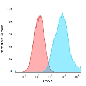
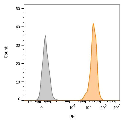
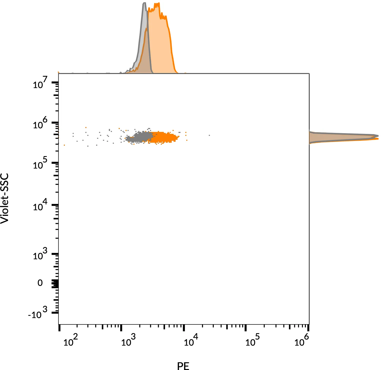
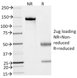
Exosome staining (verified) | Flow, surface (verified) | IF (verified) | IHC, FFPE (verified) | WB (published)Validated Applications:
FC, IF, IHC, FFPE, WBField of Research:
Cancer, Cell adhesion, Exosomes/EVsPositive Control:
HT29 cells or breast tumorConcentration:
1 mg/mLBuffer:
PBS, no BSA, no azideMolecular Weight:
40-43 kDaAdditionnal Information:
Higher concentration may be required for direct detection using primary antibody conjugates than for indirect detection with secondary antibody|Immunofluorescence: 1-2 ug/mL|Does not react with rat or ferret; others not known|Immunohistology formalin-fixed 0.5-1 ug/mL|Staining of formalin-fixed tissues requires boiling tissue sections in 10 mM citrate buffer, pH 6.0, for 10-20 min followed by cooling at RT for 20 minutes|Flow Cytometry 0.5-1 ug/million cells/0.1 mL|Western blotting 0.5-1 ug/mL|Optimal dilution for a specific application should be determined by userReferences & Citations:
Note: References for this clone sold by other suppliers may be listed for expected applications. Mol Cell Biol 21(7): 2570-2580. (western; epitope mapping)Shipping Conditions:
Room temperatureStorage Conditions:
-35°C to -5°C ; Stable at room temperature or 37°C (98°F) for 7 days.Shelf Life:
2 yearsCAS Number:
9007-83-4
DATASHEET Document
View DocumentMSDS Document
View Document