Anti-CD106 / VCAM1(VCAM1/843)
CAT:
37-BNUB0843-100
Size:
100 µL
Price:
Ask
- Availability: 24/48H Stock Items & 2 to 6 Weeks non Stock Items.
- Dry Ice Shipment: No


Anti-CD106 / VCAM1(VCAM1/843)
Description:
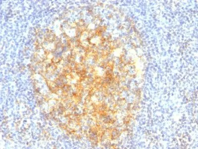
CD106 (also known as vascular cell adhesion molecule-1, or VCAM-1) is a member of the Ig superfamily of adhesion molecules. It is expressed at high levels on cytokine stimulated vascular endothelial cells, and at minimal levels on un-stimulated endothelial cells. It is also present on follicular and inter-follicular dendritic cells of lymph nodes, myoblasts, and some macrophages. CD106 serves as a ligand for leukocyte beta-1 integrin VLA-4 and mediates cell adhesion of leukocytes to activated endothelium. It plays a role in various immunological and inflammatory responses.Primary antibodies are available purified, or with a selection of fluorescent CF® Dyes and other labels. CF® Dyes offer exceptional brightness and photostability. Note: Conjugates of blue fluorescent dyes like CF®405S and CF®405M are not recommended for detecting low abundance targets, because blue dyes have lower fluorescence and can give higher non-specific background than other dye colors.Synonyms:
CD106; VCAM-1; Vascular Cell Adhesion Molecule 1; VCAM1; INCAM-100UNSPSC:
41116161UNSPSC Description:
Primary and secondary antibodies for multiple methodology immunostaining detection applicationGene Name:
VCAM1Gene ID:
7412NCBI Gene ID:
109225UniProt:
P19320Cellular Locus:
Plasma membraneHost:
MouseSpecies Reactivity:
HumanImmunogen:
Recombinant human VCAM1 proteinTarget Antigen:
CD106 | VCAM-1Clonality:
MonoclonalIsotype:
IgG1 κClone:
VCAM1/843Conjugation:
Purified, with BSASource:
AnimalApplications:
IHC, FFPE (verified)Validated Applications:
IHC, FFPEField of Research:
ImmunologyPositive Control:
Human placenta or tonsilConcentration:
0.2 mg/mLBuffer:
PBS, 0.05% BSA, 0.05% azideMolecular Weight:
~110 kDaAdditionnal Information:
Higher concentration may be required for direct detection using primary antibody conjugates than for indirect detection with secondary antibody|Immunofluorescence: 1-2 ug/mL|Immunohistology formalin-fixed 0.5-1 ug/mL|Staining of formalin-fixed tissues requires boiling tissue sections in 10 mM Tris with 1 mM EDTA, pH 9.0, for 10-20 min followed by cooling at RT for 20 minutes|Flow Cytometry 0.5-1 ug/million cells/0.1 mL|Optimal dilution for a specific application should be determined by userShipping Conditions:
Room temperatureStorage Conditions:
4°C; Stable at room temperature or 37°C (98°F) for 7 days.Shelf Life:
2 yearsCAS Number:
9007-83-4
DATASHEET Document
View DocumentMSDS Document
View Document