Anti-EpCAM / CD326 (Epithelial Marker) (EGP40/2041R)
CAT:
37-BNUM2041-50
Size:
50 µL
Price:
Ask
- Availability: 24/48H Stock Items & 2 to 6 Weeks non Stock Items.
- Dry Ice Shipment: No


Anti-EpCAM / CD326 (Epithelial Marker) (EGP40/2041R)
Description:
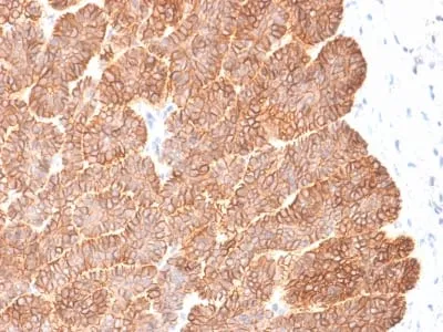
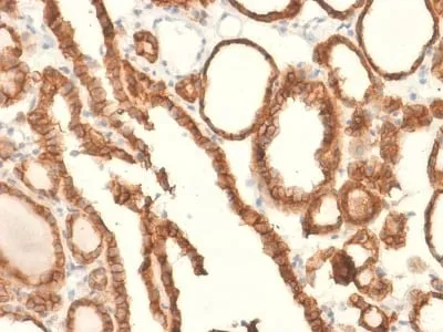
Epitope of this MAb is mapped between aa 202-212 of EGP40, which is a 40-43 kDa transmembrane epithelial glycoprotein, also identified as epithelial specific antigen (ESA), or epithelial cellular adhesion molecule (Ep-CAM). It is expressed on baso-lateral cell surface in most simple epithelia and a vast majority of carcinomas. Antibody to Ep-CAM has been used to distinguish adenocarcinoma from pleural mesothelioma and hepatocellular carcinoma. This antibody is also useful in distinguishing serous carcinomas of the ovary from mesothelioma._x000D_ _x000D_ Primary antibodies are available purified, or with a selection of fluorescent CF® Dyes and other labels. CF® Dyes offer exceptional brightness and photostability. Note: Conjugates of blue fluorescent dyes like CF®405S and CF®405M are not recommended for detecting low abundance targets, because blue dyes have lower fluorescence and can give higher non-specific background than other dye colors._x000D_ _x000D_Synonyms:
Adenocarcinoma-associated Antigen; Cell Surface Glycoprotein Trop-1; EGP2; EGP314; EGP40; Epithelial Cell Adhesion Molecule; Epithelial Glycoprotein 314; ESA; KSA; TACD1; TROP1; Tumor-associated Calcium Signal Transducer 1 (TACSTD1)UNSPSC:
41116161UNSPSC Description:
Primary and secondary antibodies for multiple methodology immunostaining detection applicationGene Name:
TACSTD1Gene ID:
4072NCBI Gene ID:
542050UniProt:
P16422Cellular Locus:
Plasma membraneHost:
RabbitSpecies Reactivity:
HumanImmunogen:
Recombinant human EpCAM fragment from extracellular domain (around aa100-224) (exact sequence is proprietary)Target Antigen:
CD326 | Ep-CAMClonality:
Recombinant MonoclonalIsotype:
IgGClone:
EGP40/2041RConjugation:
Purified, BSA-freeDisease:
TumorSource:
AnimalApplications:
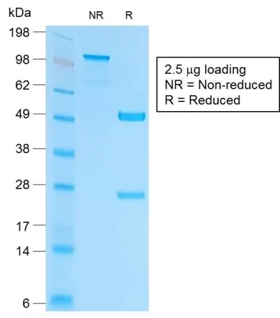
IHC, FFPE (verified) | WB (verified)Validated Applications:
IHC, FFPE, WBField of Research:
Cancer, Cell adhesionPositive Control:
HT29 cells or breast tumorConcentration:
1 mg/mLBuffer:
PBS, no BSA, no azideMolecular Weight:
40-43 kDaAdditionnal Information:
Higher concentration may be required for direct detection using primary antibody conjugates than for indirect detection with secondary antibody|Immunohistology (formalin): 0.5-1 ug/mL for 30 minutes at RT|Staining of formalin-fixed tissues requires boiling tissue sections in 10 mM citrate buffer, pH 6.0, for 10-20 minutes followed by cooling at RT for 20 minutes|Optimal dilution for a specific application should be determined by userShipping Conditions:
Room temperatureStorage Conditions:
-35°C to -5°C ; Stable at room temperature or 37°C (98°F) for 7 days.Shelf Life:
2 yearsCAS Number:
9007-83-4
DATASHEET Document
View DocumentMSDS Document
View Document