CFSE Green Fluorescent Stain - for cell labelling and proliferation studies
CAT:
436-6162
Size:
50 µg
Price:
Ask
- Availability: 24/48H Stock Items & 2 to 6 Weeks non Stock Items.
- Dry Ice Shipment: No


CFSE Green Fluorescent Stain - for cell labelling and proliferation studies
Description:
CFSE is a fluorogenic reagent which is frequently used in cell labeling and cell proliferation procedures. Non-fluorescent and moderately hydrophobic in its di-esterified form, the dye easily penetrates lipid bi-layers of living cells and is quickly converted to the amine-reactive, green fluorescent form (CFSE) upon cleavage of the acetate groups by intracellular esterases. This highly fluorescent form is membrane-impermeant and is retained on and within the cells, while any excess probe is easily washed away in subsequent wash steps. Because CFSE forms a strong bond inside the cell, it is retained within the cell indefinitely and is inherited by daughter cells. It will not be incorporated into adjacent cells.Label:
ICTCell Type:
Jurkat; K562Type:
Cell ViabiityDetection Method:
Flow cytometry, Fluorescence microscopeWavelength:
492 nm / 520-540 nmAssay Protocol:
1. Reconstitute the vial of 7-AAD with 0.26 mL DMSO to create a stock concentrate at 1 mg/mL. Mix by swirling or tilting the vial, allowing the DMSO to travel around the base of the amber vial until completely dissolved. At room temperature, the reagent should be dissolved within a few minutes forming a red solution. 2. If storing the stock concentrate for future use, prepare small aliquots (50 µL) to avoid freeze-thaw cycles. The stock concentrate will be stable for 6 months when protected from light and stored at or below -20°C., 3. Expose cells to the experimental conditions., 4. Create 2 control tubes: untreated viable cells and untreated killed cells. These cells will be stained with 7-AAD to compensate the flow cytometer to ensure that dead cells shift along the FL3 axis. These controls will also determine the level of spontaneous cell death that normally occurs within the cell line when compared with the treated cells., 5. Stain cells at a final concentration of 5 µg/mL of 7-AAD in the cell culture. This can be accomplished by pipetting the stock solution directly into the cell suspension at 1:200; e.g., add 2 µL stock to 400 µL cell suspension. This can also be accomplished by diluting the stock concentrate 1:10 to form the working solution, and then pipetting the working solution into the cells at 1:20. For example, add 50 µL 7-AAD stock concentrate into 450 µL PBS or sterile media. Mix by inverting or vortexing the vial at room temperature. Store on ice up to 2 hours. Then add the working solution to the cell suspension at approximately 1:20; e.g., put 25 µL diluted 7-AAD working solution into 475 µL cell suspension., 6. Incubate 10-30 minutes on ice., 7. If desired, wash cells twice with PBS and fix in 1% paraformaldehyde., 8. Analyze with a flow cytometer: excitation at 546 nm; emission at 647 nm in FL3. Dead cells with compromised membranes will appear red.Shipping Conditions:
Ships overnight (domestic), International Priority ShippingStorage Temperature:
-20°CCalculated Molecular Weight:
557.47Cellular Imaging & Detection:
Cellular ImagingTarget Description:
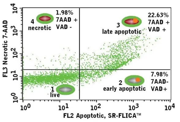
CFSE is a green fluorogenic reagent that binds to intracellular molecules. It is often used for cell proliferation and cytotoxicity studies. CFSE diffuses into the cell and covalently binds to primary amino groups present on intracellular molecules. Intracellular esterases quickly cleave the acetate groups from the dye thus converting it to the fluorescent form. Any unbound reagent diffuses back out of the cell. Because CFSE forms a strong bond inside the cell, it is retained within the cell indefinitely and is inherited by daughter cells. It will not be incorporated into adjacent cells. CFSE is supplied as a concentrated lyophilized powder at 0.05 mg. Reconstitute it with 200 µL DMSO to yield a stock concentrate at 2500X (0.25 mg/mL). Dilute it 1:250 in PBS to form the 10X working solution, and then add it to cells at 1:10 (a final concentration of 0.1 µg/mL). Analyze with a fluorescence microscope or flow cytometer with a 488 nm blue argon excitation laser. CFSE exhibits green fluorescence in the FL1 region: excitation at 492 nm and emission at 520-540 nm (Figures 1-3). As CFSE is detected in the green range, it is optimal for use in dual- staining studies with other fluorescent reagents, such as PI (catalog #638), 7-AAD (catalog #6163), and SR-FLICA® reagents with minimal spectral overlap. It can be used with our SR-FLICA® poly caspases inhibitor reagent (catalog #917) to identify apoptotic cells in the cell sample (Figure 3).